Adult Osteopathy

Diastasis Recti

During pregnancy, the abdominal muscles need to adapt to make room for the growing uterus. Diastasis recti (DRA) is when the muscles on the front of the abdomen separate (as they should), but do not come back together as well as expected after pregnancy. Please note that recovery after giving birth is a slow process, and although guidelines for DRA are limited, we do not recommend assessing, diagnosing, or attempting to treat DRA before 6 weeks postpartum.

Anatomy of Diastasis Recti

The muscles on the anterior abdominal wall are joined in the middle by the linea alba. This is a long piece of tissue, not dissimilar to a tendon, that runs from the bottom of the sternum (breastbone) to the pelvis. It is able to stretch to accommodate a growing baby (or other mass, including fat), but it is not contractile tissue like muscle. This means that it does not shrink back quickly once the need for expansion is removed.

Diagnosing Diastasis Recti

Frustratingly, there is little in the way of guidance for diagnosing or managing DRA. Good resources urge practitioners to take the depth of separation into account, as well as the width and length. As mentioned above, this should not be assessed before 6 weeks post partum, as diagnosing muscle separation before the body has had a chance to heal is not useful.

When you are past the 6 week mark, your osteopath can help you to test for a diastasis, or you can do it yourself. In clinic this will be done with you laying on your back on the plinth, or you can do it at home on your bed or the floor:

  1. Laying on your back with your knees bent, place your fingers gently on the centre of your abdomen. A diastasis often occurs around the belly button, but it can be anywhere from the bottom of the sternum to the pelvis. You may need to reassess with your hands in a different place to be sure where your diastasis is.

  2. Raise your head up as if looking down at your tummy, so that the abdominal muscles engage.

  3. If you feel a gap under your fingers with the muscles engaged either side, you may have a diastasis.

General guidance suggests that a gap smaller than two finger widths does not warrant a diagnosis. You may still benefit from some rehabilitative exercises, especially if the gap is deep.

Effects Elsewhere

We often think of core “weakness” as a factor in lower back pain. Research is actually quite inconclusive, and a diastasis is not always a sign of muscle weakness anyway.

There is a stronger link between diastasis and hernia, which makes sense as there is less tissue for the intestines to push against before they can form a hernia here. This is more reason to avoid high intra-abdominal pressure, as you would get from exercises like crunches. If you feel a lump that you suspect is a hernia, you should get it checked to be sure. Note that the strangulation of a hernia, or a hernia that is significant enough to cause a bowel blockage are red flags. Severe abdominal pain or a reduction in bowel movement should be investigated urgently if you have a hernia.

Management Strategies

The best approach is focused on strengthening exercises. This is a slow process, as the stretched tissue itself is not a muscle, but it may be influenced by the muscles that it attaches to. Core exercises are good, but you need to address lateral and rotational movements as well as the usual flexion- and crunch type exercises are best avoided. Your osteopath can work with you to devise an exercise plan that suits you best, using exercises such as planks, side planks, and other variations. Don’t underestimate the demand of day to day activity on your core either; for example walking incorporates the movements mentioned above with minimal strain.

Click here to make an appointment for your diastasis in Naas
Cervicogenic Headaches (From the Neck)

Headaches can be notoriously difficult to find a root cause for. Some headaches come from structures in the neck, which can be relatively easy to test for. In many cases, treatment can be quite straight forward too.

Identifying Cervicogenic Headaches

Cervicogenic just means “from the neck”. Symptoms can be quite similar to tension-type headaches (TTHs), for which treatment is different. The main difference between the two is that TTHs are considered “primary headaches”. This means that they are not caused by another condition. Cervicogenic headaches can be diagnosed whenever a headache can be attributed to a known issue in the neck, either by clinical diagnosis or imaging. Alternatively, they can be diagnosed if two of the following points are true:

  1. A headache develops alongside the development of a neck problem

  2. The headache improves or resolves when the neck problem does the same

  3. Neck movement is limited, and some movements aggravate the headache

  4. Diagnostic blocks (such as nerve blocks) effectively remove headache symptoms

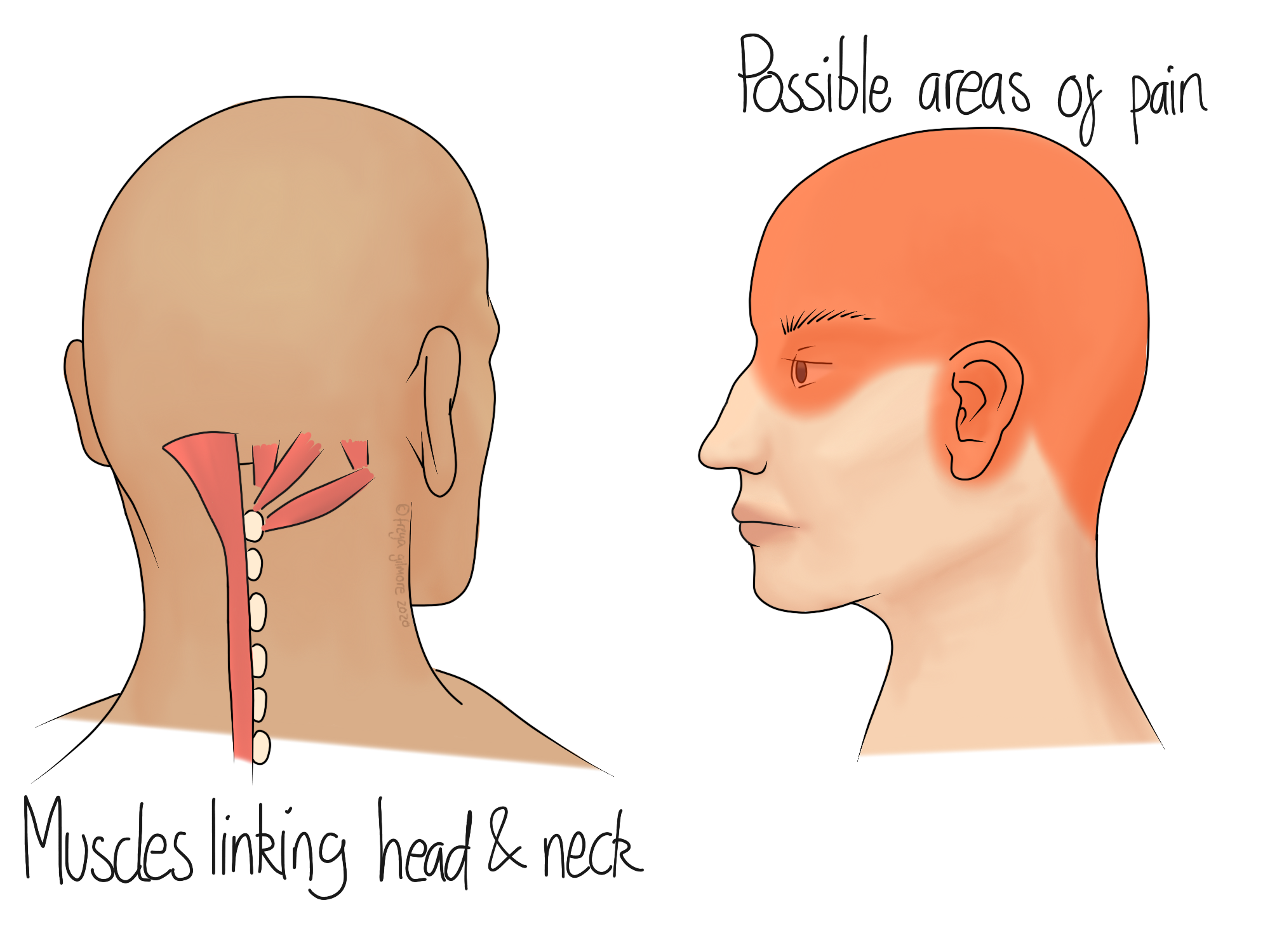
Symptoms can be anywhere in the red area illustrated. Despite covering the eye, they will not cause visual disturbances. Dizziness and nausea are not typical symptoms either. Headaches with these additional symptoms may indicate migraine, for which we may still be able to help. Pain may feel like it radiates up from the neck and over the top or side of the head. If the cause is a tight muscle in the neck, you might find that a warm compress on the neck, or resting the head help. For other causes, a cool compress may be more beneficial. Your osteopath can advise the more appropriate for you.

Tension Type Headaches

TTHs have the following symptoms:

  1. They cause pain on both sides of the head

  2. The pain can be described as pressing, or tightening, but not pulsating

  3. Intensity of the pain is mild to moderate

  4. Typical physical activity, like walking or going up stairs, does not aggravate symptoms

There can be a lot of variation between TTHs, and a single headache can last anywhere from 30 minutes to over a week. In contrast, cervicogenic headaches usually last less than a day, but may be frequent.

Managing Cervicogenic Headaches

Osteopaths are qualified to help with these kinds of headache. We will assess the neck, and usually the upper back too, to determine what structures may be having an effect. Often the muscles and joints of the neck are overworked by restricted areas further down the spine. This can be particularly evident with underlying conditions like osteoarthritis. When arthritis affects the hips, it can encourage you into a forward posture, for which the neck has to compensate. In cases like these, we might find that improving hip and lower back function takes the stress off the neck, and improves the headaches.

Whatever the underlying cause, or causes of your headaches, your osteopath will work to identify as many as possible. Treatment is gentle and drug free, using massage, stretching, and joint manipulation techniques to improve function throughout the body. We aim to take the burden off the overworked areas and get your whole body moving more comfortably and efficiently.

Click here to make an appointment in Naas for your headaches

Tennis Elbow

Tennis elbow is the common name for lateral epicondylitis. The epicodyle is part of the humerus (upper arm bone) that sits close to the elbow. It is where the muscles attach that control wrist movement. The "-itis" in the name refers to the inflammation that occurs in the condition, which is focused around the connection between the muscle and the bone.

"Tennis" Elbow

Despite the name, the condition can develop without the patient ever having touched a tennis racket. The muscles that become problematic are the same that are used in a backhand swing, but of course this is not all they are used for. Moving a heavy kettle or pan of water can put demand on the same muscles, known as the wrist extensors.

Golfer's Elbow

The opposite side of the arm can be affected in the same way. This is medial epicondylitis, colloquially known as "golfer's elbow". As before, you can develop this condition without playing the sport it is named after. Wrist flexion could be overused when carrying large shopping bags that would drag on the floor unless you engage your wrist. Even carrying a child for a long time could require extra work from these muscles.

The Process

Rather than tennis elbow being an example of a tight muscle, it's a process beyond that. If the muscle is tight or strained enough for long enough, it can begin to irritate the join between muscle and bone. This causes inflammation, at which point symptoms can change. Whereas before there might have just been some local aching or tenderness in the muscle, the pain may become more deep and persistent. Inflammation often responds well to a cool compress whereas simple muscle tightness often prefers heat. Your osteopath will ask if you've found anything that makes the symptoms better or worse, so these factors can be quite telling.

Osteopathy and Tennis Elbow

Your osteopath will take a holistic approach at managing your pain. We want to get to the bottom of why the condition developed in the first place, as well as providing relief. Treatment itself might involve massage techniques to the affected muscles, as well as mobilising techniques for the elbow and wrist joints. Self management can be really important here, especially if the cause is identified as a factor that could be modified.

When epicondylitis is actually related to racket sports, sometimes it can respond to a simple change in the grip. If you racket's grip is skinny, it asks the affected muscles to work harder just to hold on, predisposing the problem before the game has even started. Wrapping the handle with an additional roll of grip tape might ease the load enough for you to continue playing while getting better.

If your symptoms are worse in the morning, it might be related to your sleeping position. We don't often recommend braces to immobilise joints, but in this case it might be appropriate. An off-the-peg wrist brace can sometimes be a good way to prevent tension in the affected muscles overnight, especially if you find that you sleep in a curled up position. You won't be wearing the brace while awake, as general movement is key to maintaining strength and mobility. It also helps to move the inflammation along.

Click here to make an appointment for your tennis elbow in Naas

Chronic Pain

When pain persists for over three months, it is considered chronic. When it is present for a long period like this, it can affect the patient biologically, psychologically, and socially.

Chronic pain is complex, and influenced by a number of factors. The image above shows a few. Physical health plays a role, affected by sleep, smoking, and diet. Psychology is important too- if someone is told that their bones are “crumbling” it will affect the way they move and trust their body. Some medications can increase sensitivity to pain, and ongoing insurance claims can genuinely affect symptoms.

How Chronic Pain Works

You can think of pain as a protector- it is there for a reason. It can be overprotective though, becoming hypersensitive. This can manifest as more intense pain than normal, or pain in response to things that shouldn’t cause pain.

The hypersensitivity can be blamed on the nervous system. The area in pain is not to blame. But the nervous system is influenced by so many different things, it can be hard to make changes on your own. A doctor may prescribe painkillers to help you get on top of things, but patients often don’t want to take them for too long. Side effects can be unpleasant, or they may be concerned that the cause has not been addressed. Pharmacology can work really well alongside therapies like osteopathy to start to unpick the problem.

Ongoing Stimulus

Chronic pain can be caused by a condition, such as osteoarthritis, that continues to produce pain. This is known as “secondary chronic pain”. Although this can be associated with the hypersensitivity mentioned above, it is not always. This kind of pain is easier to manage, as fixing or improving the cause will reduce the pain.

Primary Chronic Pain

If your pain cannot be attributed to another condition, it is considered primary chronic pain. Fibromyalgia is one of a handful of conditions that count as a primary cause.

There is a grey area over chronic lower back pain. It can be difficult to pinpoint the exact cause of a case of lower back pain. Even MRIs are imperfect, as many people have structural changes (like bulging discs) without any symptoms. We know that chronic lower back pain does exist once the original problem has resolved, and in this case it would be considered primary. But it is not always clear if someone’s original stimulus has resolved or not.

Your osteopath has tests they can perform to help decipher the cause of your pain. If you have already had investigations like imaging and blood tests, bring your results to your appointment as they may be useful.

Osteopathy and Chronic Pain

Whether your pain is primary or secondary, we may be able to help. We mentioned above that pain is influenced by many factors, but fortunately osteopathy addresses a number too. Beyond the mechanical parts of the picture, we can help with lifestyle advice, reassurance, and education to name a few. Outcomes are better among empowered patients who understand what is happening and why. We cannot cure all chronic pain, but if we may be able to help you manage it.

Book an appointment to address your chronic pain here.

Hip and Knee Osteoarthritis

Osteoarthritis (OA) can affect any joint with cartilage inside. Two commonly affected joints are the hip and knee. You can read more about the process of osteoarthritis here.

Knee Arthritis

There are actually three joints in the knee, and any one of them can develop OA. The main joint is the big hinge joint between the thigh and shin bone. OA is an issue of cartilage, and there is plenty of it in this joint. Beyond the usual cartilage coating the bones within the joint, there are two additional pieces in each joint. These are the menisci, and their role is to support the joint by providing a better connection between the bones.

The second joint in the knee is the patellofemoral joint: where the knee cap meets the rest of the knee. This can also be subject to arthritis. The joint is quite often overworked, as it sits within the tendon for the quadriceps. If these muscles are tight, they put extra pressure on the joint and cause friction. Overuse like this is a factor in developing arthritis. Symptoms of patellofemoral arthritis can include:

  • Pain at the front of the knee

  • Pain on going up and down stairs or ramps

  • Relief when fully straightening your knee

  • Aching in the front of the thigh

The third knee joint is on the outside, where the fibula joins the rest of the knee. This joint is small and is less likely to present a problem than the others, but your osteopath will consider it if appropriate.

Hip Arthritis

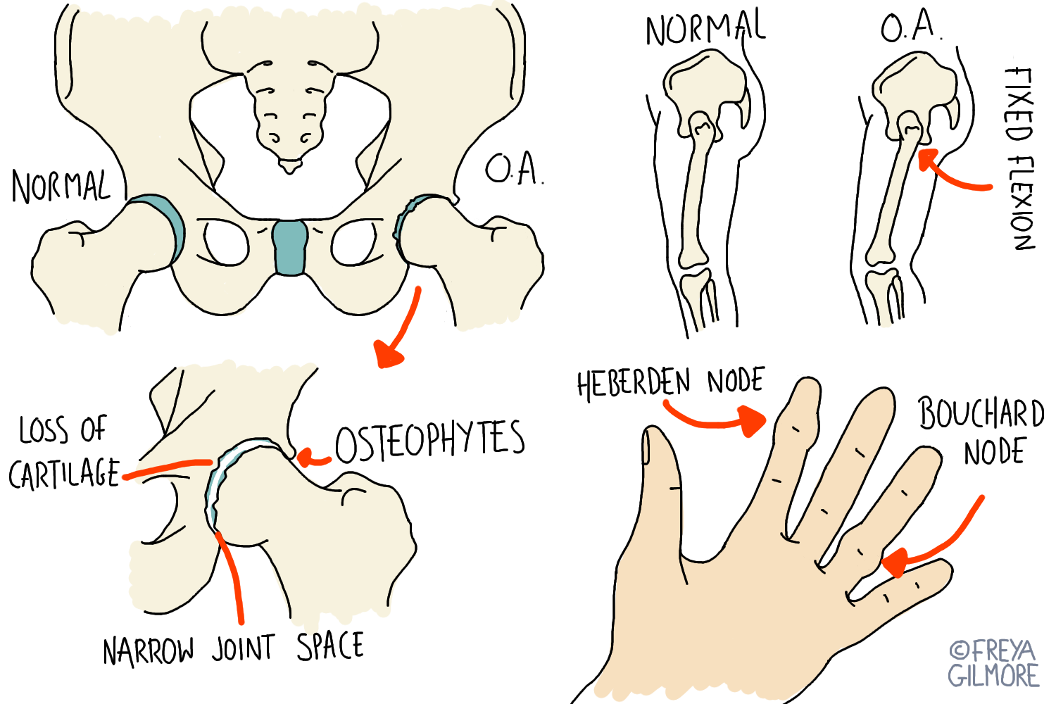
The hip can develop arthritis with very few symptoms until the late stages. Often the first sign is a loss of extension (moving your leg backwards). The body is good at adapting to this loss, and rather than challenging itself, it finds the path of least resistance. It tilts the pelvis so you can still move your leg back, but without using the hip to do so.

Hip arthritis is associated with compensation through the back and neck, which can ultimately cause the development of more arthritis in these areas.

One of the most common joints to be replaced is the hip. Contrary to popular belief, this is not the only outcome for people who develop arthritis in the joint. In the early stages, it may even be possible to reverse some of the damage if you can achieve better movement through the hip. The more progressed the condition, the more likely you are to be considered for surgery, so early action is key.

Osteopathy for Osteoarthritis

Your osteopath is qualified to help with your arthritis. We can help to improve the movement in the joint, which will allow the body to get as much nutrition as it can to the area. We will also look at any compensatory patterns that have developed elsewhere in response to the arthritis. Preventing and managing new symptoms may be realistic treatment goals for you.

Click here to make an appointment in Naas for your arthritis.

Osteoarthritis

Osteoarthritis (OA) is a condition that affects most of us eventually. We used to think of it as "wear and tear" of joints, but now we know there's also a "repair" element to it.

What happens in Osteoarthritis?

OA is a condition of cartilage. Cartilage cushions almost all of our major joints, but it has a poor blood supply. This means that it relies on nutrient exchange from surrounding fluid to stay healthy.

It's easier for cartilage to receive nutrients and flush out waste if it goes through compression and decompression. You can imagine it as a dirty sponge in clean water: pump it enough and it'll come out clean.

Early stages

During the early stages of osteoarthritis, the cartilage stops being properly pumped. This may be subconscious, and it may be the body's attempt at protecting itself from pain or injury. If this is not addressed, the cartilage begins to become unhealthy.

As the cartilage becomes more unhealthy, you may find there is more discomfort. This encourages further compensation to take pressure off the sore joint. Unfortunately, this means the cartilage is still not being pumped, and the cycle is not being broken.

The rest of the body continues to adapt, often without you noticing. If the hip is affected, your posture might change so you don't have to use the painful range of movement. This causes compensation elsewhere, leading to a hunched posture. Over time, this can also cause problems in the lower back and neck as their demand increases.

Late stages

If OA is not addressed, the cartilage continues to decline. The surface of the joint becomes more rough, and pieces of cartilage may become loose. Lost cartilage can be replaced by bone, and bone might grow around the edge of the joint, forming a "lip" visible on X-Ray.

By now, the joint is likely symptomatic. Crunchy, painful, or limited movement can all be signs of osteoarthritis. The body continues to try and protect itself, limiting movement by tightening surrounding muscles. It is not too late to seek treatment, as further restriction of movement will only speed up the degenerative process.

Beneath the cartilage, cysts can form within the bone. At this point there is only so much that can be done with manual therapy. By now, the joint is likely to be significantly painful, and a joint replacement may be the best course of action.

How can OA be treated?

The good news is that manual therapy has been proven to be effective in improving pain and mobility in arthritic joints. Thinking about how OA develops, it makes sense that hands on treatment and exercise would help.

Your osteopath can spot the early signs of osteoarthritis, possibly even before you have. In very early cases it may be possible to fully restore the health of the joint.

For most patients, treatment involves work to get the joint to a comfortable level, then exercises and longer term management to keep it happy. If there have been compensations elsewhere in the body, these can be addressed too.

Nip your osteoarthritis in the bud. Make an appointment in Naas here.

Osteopathy in Pregnancy

The body goes through changes at an unprecedented speed during pregnancy, so symptoms of lower back pain are unsurprising. However, you do not need to suffer through them.

Hormonal Causes of Back Pain

From early in the first trimester, the body begins to produce higher levels of the hormone “relaxin”. The purpose of this hormone is to increase movement within the pelvis by relaxing the ligaments around it in preparation for birth. Sometimes lower back pain can be one of the first signs of pregnancy for this reason.

Hormone levels peak again in the third trimester, at which point back pain may return or develop for the first time. If the joints of the pelvis are painful, they may be worse when:

  • turning over in bed

  • getting up from sitting

  • walking

Your osteopath cannot reduce the effects of the hormone itself, but we may be able to help your body cope better with the changes.

Although effects of relaxin may be focused to the pelvis, there are systemic changes too. Relaxin affects the permeability of blood vessels, which can cause fluid retention. This may be mild, and only recognised later on when rings no longer fit comfortably. Alternatively, it may be significant, even causing carpal tunnel syndrome.

Mechanical Causes of Back Pain

As the baby bump develops, the centre of gravity shifts forwards. In order to keep balanced, posture needs to adapt. The typical adaptation is an increase in the curve at the bottom of the back. Directly, this encourages the muscles of the lower back to shorten and tighten. We also expect to see tightness in the buttock and hamstring muscles as they work to keep you upright.


When the lower back arches more than usual, the demand on the rest of the spine changes. The upper back may become stiff, and the angle of the neck needs to change to keep the head level. Some treatment methods are inappropriate during the later stages of pregnancy, but your osteopath will adapt their plan for you. Increasing movement through the neck and upper back, as well as managing associated muscle tightness can make all the difference.

More Severe Pelvic Joint Pain

SPD stands for Symphysis Pubis Dysfunction, which can be agonising. In this condition, the combination of hormonal and mechanical changes causes irritation of the large cartilage joint at the front of the pelvis. If left unmanaged, this can impact birth itself, as birthing positions with legs apart are not recommended. Some women also require the use of crutches during the later stages of pregnancy. Symptoms may clear up spontaneously at birth, but there are things we can do to help you manage in the meantime.

Make an appointment online to manage or prevent back pain in pregnancy.